Horse Hind Leg Tendon Anatomy / Forever Horses Anatomy Of The Equine Forleg / The arterial supply to the digit and fetlock of the thoracic limb comes mainly from the median palmar artery.the median palmar artery divides in the distal fourth of the metacarpus between thesuperficial and deep digital flexor tendons and the suspensory ligament, to become the medial and lateral digital arteries.part of the deep palmar arch anastamoses with the lateral digital.
byAdmin•
0
Horse Hind Leg Tendon Anatomy / Forever Horses Anatomy Of The Equine Forleg / The arterial supply to the digit and fetlock of the thoracic limb comes mainly from the median palmar artery.the median palmar artery divides in the distal fourth of the metacarpus between thesuperficial and deep digital flexor tendons and the suspensory ligament, to become the medial and lateral digital arteries.part of the deep palmar arch anastamoses with the lateral digital.. The horse's hind limbs the top part of the hind limbs consists of three fused bones, called the ileum, ischium, and pubis. Swelling of the sdft results in a curvature of the flexor tendons in the cannon region. The limbs of the horse are structures made of many bones, joints, muscles, tendons and ligaments that support the weight of the horse's body. Tenosynovitis is the swelling of the digital sheath that surrounds the superficial and deep flexor tendons in the leg of the horse. Fat and muscle in order to visualize the lengths and angles of the bones that lie beneath.
The part of the hindquarters behind the thighs and below the root of the tail Swelling of the sdft results in a curvature of the flexor tendons in the cannon region. An example of this is when the flexor tendons of the hind leg can flex the fetlock, pastern and coffin joint, but also extend the hock joint. The horse's hind limbs the top part of the hind limbs consists of three fused bones, called the ileum, ischium, and pubis. They are joined to the spine through the sacroileac joints and allow transfer of propulsion to the hind legs.
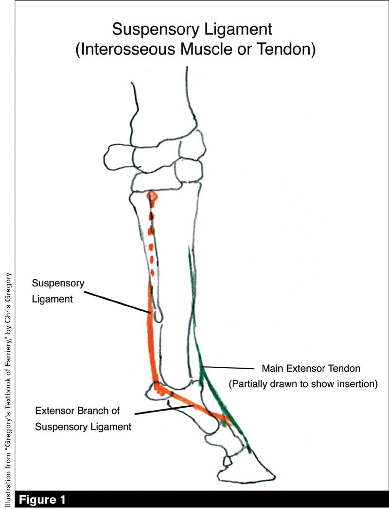
Increased Knowledge Of The Equine Anatomy Can Help Farriers Improve Hoof Care American Farriers Journal from www.americanfarriers.com The tendons extend from muscles high in the leg to the foot; Horse hind leg anatomy sectional view in this image, you will find the proximal suspensory ligament, deep digital flexor tendon, suspensory ligament, superficial distal sesamoidean ligament, deep digital flexor tendon, extensor branch of suspensory ligament in it. The anatomy of dressage horse hindquarters. The pictures will help you see which muscles you are stretching! It also includes the joints of the hip, stifle, hock, fetlock, pastern, and coffin It is the major extensor tendon of the leg. The majority of the power of movement should come from the rear legs. Gastrocnemius tendonitis in horses is a serious condition of the gastrocnemius tendon, which is the muscle that attaches the upper part of the leg to the hock.
The tendons most often damaged in performance horses are the superficial digital flexor (sdft) and the suspensory ligament (sl) which is the same as the interosseous tendon.
The horse's hind limbs the top part of the hind limbs consists of three fused bones, called the ileum, ischium, and pubis. In contrast, the deep digital flexor tendon (ddft) has a lower incidence of damage. Tenosynovitis is the swelling of the digital sheath that surrounds the superficial and deep flexor tendons in the leg of the horse. Florent david's approach, which he presented at the 2019 neaep symposium. There are two tendons that run down the back of the leg. The digital flexor tendon sheath on the back of the fetlock joint helps ease the passage of the deep digital flexor tendon past the fetlock joint. The ischium forms the point of the buttock. Horse anatomy and muscle diagrams. Muscles, tendons, and ligaments run along the long tibia and smaller fibula to the hock joint. Massage is a powerful tool in maintaining and increasing your horse. The horse leg anatomy in the rear includes the bones of the pelvis (the ilium, ischium and pubic bones), femur, tibia, fibula, metatarsus and the phalanxes. Directional terms, skeletal, and muscle introduction. These horse anatomy diagrams are a great overview and introduction to the vast study of equine anatomy.
The part of the hindquarters behind the thighs and below the root of the tail The horse's hind limbs the top part of the hind limbs consists of three fused bones, called the ileum, ischium, and pubis. Tendon and musculature) is not lame, but rapidly learns to compensate by flicking the lower limb using the carpal or tarsal extensor units. The pictures will help you see which muscles you are stretching! All images are contained in the stretch your horse app for free!
The Action Of Muscles from static.wixstatic.com The body of the horse, enclosing the rib cage and the major internal organs; The ischium forms the point of the buttock. Here's a look at dr. Massage is a powerful tool in maintaining and increasing your horse. It also includes the joints of the hip, stifle, hock, fetlock, pastern, and coffin. A horse to may pull its weight off its hind leg early thus weighting and stressing the opposite shoulder. The limbs of the horse are structures made of many bones, joints, muscles, tendons and ligaments that support the weight of the horse's body. Appreciate the strain the tendons and ligaments have to absorb when landing from a.
In the hind limb, the ddft originates from two areas of the tibia and also inserts on the coffin bone.
The horse leg anatomy in the rear includes the bones of the pelvis (the ilium, ischium and pubic bones), femur, tibia, fibula, metatarsus and the phalanxes. Fat and muscle in order to visualize the lengths and angles of the bones that lie beneath. If the tendon is ruptured, the horse’s leg will collapse when putting weight on it, causing a major case of lameness. When either of these tendons swell it causes the leg to look bowed. the bow can appear anywhere from the knee or hock to the pastern region. Anatomy of front limb lower horse leg. That way if you need to talk to a vet, or do a correct drawing, you'll have a solid foundation. A horse to may pull its weight off its hind leg early thus weighting and stressing the opposite shoulder. In the hind limb, the ddft originates from two areas of the tibia and also inserts on the coffin bone. This page contains color coded pictures of the horse's hind end, deep and superficial muscles. The part of the hindquarters behind the thighs and below the root of the tail The limbs play a major role in the movement of the horse, with the legs performing the functions of absorbing impact, bearing weight and providing thrust. A guided tour scott j. The longissimus dorsi, is the main muscle in the horse's back and underneath the saddle.
Muscle or the muscle group increases the risk of injury, causes a shortened forward stride, a resistance to lateral work and joint discomfort in the hindquarters. It is the major extensor tendon of the leg. Muscles, tendons, and ligaments run along the long tibia and smaller fibula to the hock joint. The horse's hind limbs the top part of the hind limbs consists of three fused bones, called the ileum, ischium, and pubis. It also includes the joints of the hip, stifle, hock, fetlock, pastern, and coffin.
Suspensory Desmitis In Horses Musculoskeletal System Merck Veterinary Manual from www.merckvetmanual.com When either of these tendons swell it causes the leg to look bowed. the bow can appear anywhere from the knee or hock to the pastern region. This page contains color coded pictures of the horse's hind end, deep and superficial muscles. There are four classifications of tenosynovitis in horses. The small bone that forms the point of the hock is actually similar to the human heel bone. Tendon and musculature) is not lame, but rapidly learns to compensate by flicking the lower limb using the carpal or tarsal extensor units. The majority of the power of movement should come from the rear legs. They are joined to the spine through the sacroileac joints and allow transfer of propulsion to the hind legs. Horse rear legs the horse leg anatomy in the rear includes the bones of the pelvis (the ilium, ischium and pubic bones), femur, tibia, fibula, metatarsus and the phalanxes.
The one nearest to the skin is the superficial digital flexor tendon and the one nearest to the cannon bone is the deep digital flexor tendon.
The limbs of the horse are structures made of many bones, joints, muscles, tendons and ligaments that support the weight of the horse's body. The longissimus dorsi, is the main muscle in the horse's back and underneath the saddle. In a horse, however, some of the tendons can flex multiple joints and extend others. In contrast, the deep digital flexor tendon (ddft) has a lower incidence of damage. The ischium forms the point of the buttock. There are four classifications of tenosynovitis in horses. The anatomy of dressage horse hindquarters. Our latest youtube film is ready to run. The tendons most often damaged in performance horses are the superficial digital flexor (sdft) and the suspensory ligament (sl) which is the same as the interosseous tendon. What is a bowed tendon? Massage is a powerful tool in maintaining and increasing your horse. The area where the saddle sits, beginning at the end of the withers, extending to the last thoracic vertebrae (colloquially includes the loin or coupling, though technically incorrect usage); Tenosynovitis is the swelling of the digital sheath that surrounds the superficial and deep flexor tendons in the leg of the horse.
The tendons are named according to whether the joints they attach to flex or extend leg tendon anatomy. The ischium forms the point of the buttock.